Dark-field Microscopy Is Often Used to Detect Which Bacterial Shape
Dark-field microscopy describes microscopy methods in both light and electron microscopy which exclude the unscattered beam from the image. The strong brilliance of the needle-shaped MSU crystals is clearly distinguished in the dark field.
Dark Field Microscopy Principle And Uses Microbe Online
All four pathogenic treponemes belong to the Spirochaetaceae family of bacteria named for their spherical shape when examined under darkfield microscope.

. It allows the visualization of live bacteria and distinguishes some structure rods curved rods spirals or cocci and movement. The object with a cone of light is the most essential part of the dark-ground microscope. Specimen in dark field.
What would happen if a microbiology technician used an old bacterial culture 24 hr to make the slide. As a result the field around the specimen is generally dark. To maximize the scattered light-gathering power of.
Fluorescence microscopy is most often used to detect specific proteins or other molecules in cells and tissues. A very powerful and widely used technique is to couple fluorescent dyes to antibody molecules which then serve as highly specific and versatile staining reagents that bind selectively to the particular macromolecules they recognize. The causative agent of syphilis is a spiral-shaped organism.
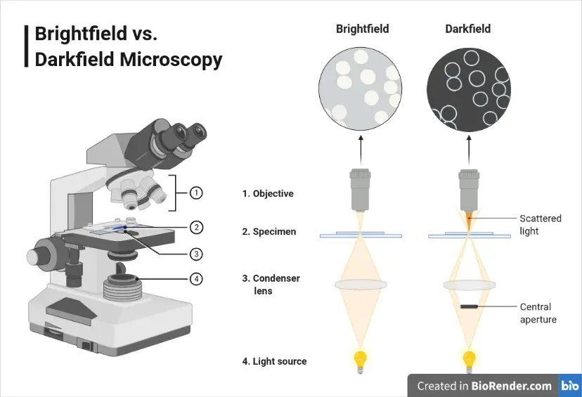
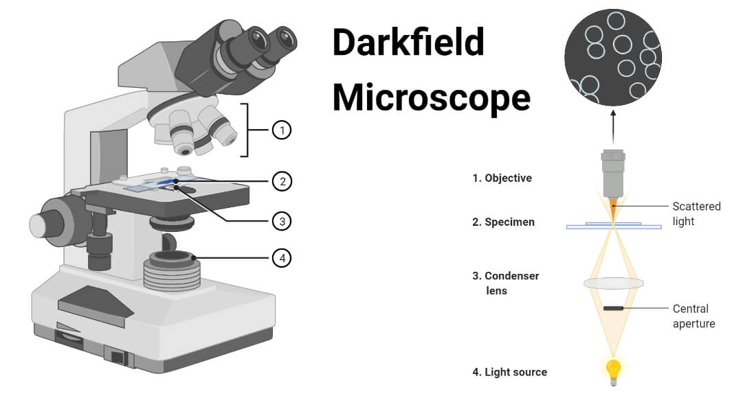
In optical microscopes a darkfield condenser lens must be used which directs a cone of light away from the objective lens. Bright bacteria on a bright background. Which of the following would NOT negatively affect the shape of an organisms growth curve.
Dark-field DF microscopy has seen relatively little use in clinical microscopy applications 1113. Dark field microscopy and Enderlein therapy. Write down your answers and then click at bottom of page for the correct answers What would you see using a dark-field microscope on bacteria that transmit light without reflecting it into the objective lens.
In 1906 immunologist Karl Landsteiner along with colleague Viktor Mucha introduced dark-field microscopy as a means of detecting the presence of syphilitic treponemes spiral-shaped bacteria in infected specimens. Spirochetes are so narrow that their shape cannot be fully resolved by light microscopy. Cocoid a coccus is any bacterium that has a spherical ovoid or generally round shape.
This is already noticeable at 200 which allows the examination of a larger microscope field and faster detection. Captured on the surface were then detected by dark field microscopy and statistical analy sis was performed on the recorded image for the counti ng of the bacteria captured by antibodies. In the beginning there was dark-field microscopy.
Dark-field microscopy has many applications in microbiology. Dark-field microscopy is used to detect spirochetes such as Treponema pallidum the causative agent of syphilis. Aspirochete bflagella ccocci.
It might be worthwhile for beginners to look at 200 and even at 100 after having detected the crystals at 400. Dark-field microscopy involves obtaining a scraping of the sore placing it. The dark-ground microscopy makes use of the dark-ground microscope a special type of compound light microscope.
Dark-field microscopy is often used to detect which bacterial shape. Dark-field microscopy is often used to detect which bacterial shape. DF has been used to image bacterial flagella 14 and is an established.
Dark bacteria on a bright background. But whereas most doctors just use darkfield microscopy to detect spirochete bacteria eg Lyme disease syphilis or yaws LBA advocates say you can use the procedure to identify a host of. The darkfield microscope is based on the theory of pleomorphism.
Dark Field Microscopy Dark field microscopy is a technique used in the clinical from BIO MISC at Emilio Aguinaldo College. The dark-field condenser with a central circular stop which illuminates. Spirochete Which type of microscopy exploits small differences in refractive index between the.
This is why the term Enderlein therapy is often used in connection with dark field microscopy. Human pathogenic treponemes measure 615 µm in length and 02 µm in diameter enveloped by a cytoplasmic membrane and a loosely associated outer membrane. The chip is slightly larger than a postage stamp and as thin as a credit card.
Bacilliform bacteria are also often simply called rods when the bacteriologic context is clear. This organism is classified as a. It is one of the three distinct bacterial shapes the other two being bacillus and spiral-shaped cells.
Phase contrast microscopy converts phase shifts in the light passing through the transparent specimen into brightness changes that are. The dark field microscopy method was developed in the 1920s by zoologist and bacteriologist Prof. Rbc red blood cell.
When placed on a microscopes stage the chip emits a hollow cone of light that can be used to generate detailed dark-field images of algae bacteria and similarly translucent tiny objects. Dark-field microscopy is often used to detect which bacterial shape. Dark field microscopy which uses an illumination technique to enhance contrast in unstained specimens producing bright objects on a dark background can be used to detect spirochetes and Leptospira but is not routinely used.
The condenser contains an opaque central disk that blocks some transmitted light allowing visualization of the sample.
Darkfield Microscope Definition Principle Uses Diagram

3 3a Dark Field Microscopy Biology Libretexts
Dark Field Microscopy Of Bacterial Colony 31 Download Scientific Diagram
No comments for "Dark-field Microscopy Is Often Used to Detect Which Bacterial Shape"
Post a Comment